Photoacoustic Imaging - EMBS Chapter Presentation at the 2018 Rochester Section Joint Chapters Meeting

The IEEE EMBS Rochester Chapter has a speaker presentation at the Section's joint chapters meeting (JCM) on March 28th, 2018. The technical sessions are free to attend for IEEE members. Reservations are required to attend the dinner and keynote presentation. Please find details and register for the JCM at:
https://events.vtools.ieee.org/m/157596
Date and Time
Location
Hosts
Registration
-
 Add Event to Calendar
- Louise Slaughter Hall
- 78 Rochester Institute of Technology
- Rochester, New York
- United States 14623
- Building: RIT Center for Integrated Manufacturing Studies Conference Center
- Room Number: Building 78
Speakers
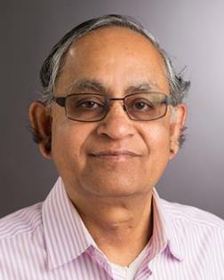
 Dr. Navalgund Rao of Rochester Institute of Technology
PHOTOACOUSTIC IMAGING: OUR EXPERIENCES WITH TECHNOLOGY DEVELOPMENT AND APPLICATION FOR CANCER DIAGNOSIS
Photoacoustic Imaging is a new promising medical imaging modality that is mapping its path from research to clinical applications. It is a hybrid technique, where light goes into the body and ultrasound comes out. Therefore it combines the superior optical absorption based oxy and de-oxy hemoglobin (blood) contrast with good spatial resolution afforded by ultrasound for cancer diagnosis. For volumetric Photoacoustic imaging, the traditional method of computed tomography based image reconstruction is expensive, cumbersome and time consuming. We have developed an innovative acoustic lens based imaging camera system that addresses many of these challenges. Alongside the technology development, we have performed ex-vivo imaging of over 100 freshly excised human prostate, thyroid and kidney specimens. With statistical pattern recognition and convolution neural network based analysis of the multispectral photoacoustic imaging data, we are able to demonstrate superiority in differentiating malignant cancer region from normal and benign tissue, when compared to state of the art clinical ultrasound evaluation of thyroid and prostate glands.
Biography:
Recently retired as a tenured Professor from the Center for Imaging Science at Rochester Institute of Technology, Dr. Rao is now working as a research professor at RIT and also holds an adjunct appointment in Imaging Sciences department, University of Rochester Medical center. He has been primarily involved in graduate teaching and research for the past 25 years. His research interests are in Physics and engineering of medical imaging, digital image and signal processing, with emphasis on ultrasound imaging. Since 2006, he has worked in collaboration with Dr. Vikram Dogra, MD and Dr. Wayne Knox at U of R. Development of Photoacoustic Imaging technology and its application for thyroid, prostate and breast cancer disease management has been the major thrust of his research for the past decade.
Address:Center for Imaging Science, Rochester Institute of Technology, New York, United States
Agenda
CHAPTER TECHNICAL PRESENTATIONS SESSION II (5:30 – 6:25 PM):
IEEE Engineering in Medicine and Biology Society (Room: SLA 2130)
Photoacoustic Imaging: Technology Development and Application for Cancer Diagnosis, Navalgund Rao, RIT

