Tissue Cytometry, Cell Cytometry & Small Animal Imaging technologies by PerkinElmer
Indistrial talk Organised by IEEE EMBS SB Chapter, IIT Kharagpur
Date and Time
Location
Hosts
Registration
- Date: 31 Jul 2018
- Time: 11:30 AM UTC to 12:30 PM UTC
-
 Add Event to Calendar
- Old SMST Seminar Room
- Opposite Kalidas Auditorium, IIT Kharagpur
- Kharagpur, West Bengal
- India 721302
- Click here for Map
Speakers
Dr Shahzada Asad of PerkinElmer (India) Pvt. Ltd.
Tissue Cytometry, Cell Cytometry & Small Animal Imaging technologies
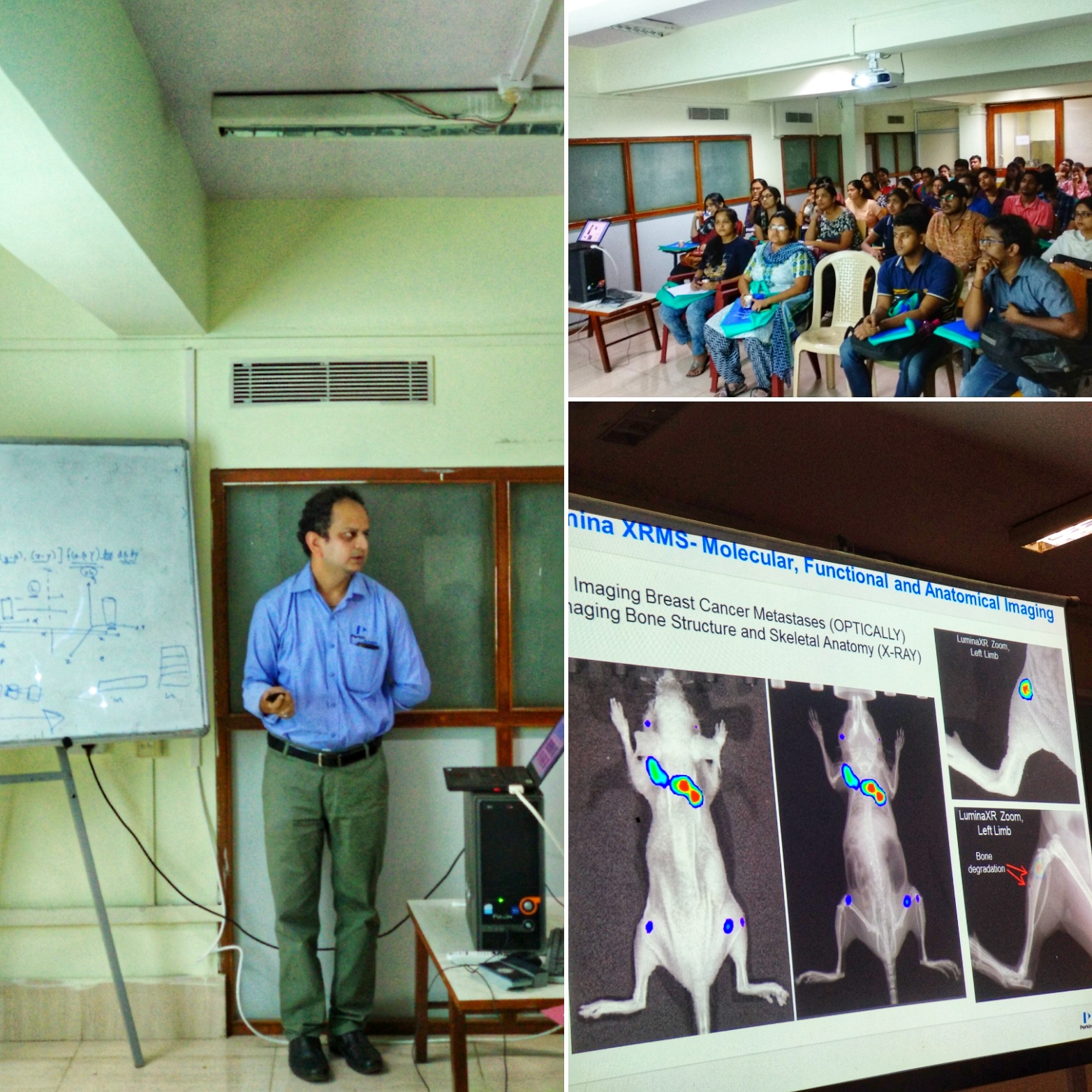
In vivo or Non invasive imaging have become indispensable research tools in many clinical and basic research laboratories, Pre-clinical imaging techniques including in vivo, ex vivo enables the understanding of disease from cellular to whole animal models, and are used in the translation to and development of therapies in clinical-relevant models. Optical imaging methods utilize fluorescence, reflectance, Bioluminescence & X-Ray contrast agents as a source of contrast. These applications are based on genetic engineering advances and improved imaging technologies. PerkinElmer offers Fluorescence & Bioluminescence, Radiolabeled Imaging Platforms for in vivo and preclinical imaging research, where user can perform 2D, X-Ray, 3D Tomography & Computed Tomography imaging for Preclinical screening.
PerkinElmer IVIS is an integrative platform that combines the full suite of optical features including Spectral Unmixing, 2D and 3D quantitative bioluminescence and fluorescence with fast and low dose CT imaging. The simple user interface along with automated co-registration, advanced visualization and analysis tools are driven by PerkinElmer’s market leading Living Image® software. The IVIS enables longitudinal workflows to characterize disease progression and therapeutic effect throughout the complete experimental time frame with both quantitative CT and optical reconstructions. Fast imaging and the ability to image multiple animals offers the throughput required to scan large cohorts of animals quickly and draw sound conclusions from your experimental data.
High-content screening (HCS) is used in biological research and drug discovery to identify substances such as small molecules, peptides, or RNAi that alter the phenotype of a cell in a desired manner. Hence high content screening is a type of phenotypic screen conducted in cells. Phenotypic changes may include increases or decrease in cellular product, High content screening includes any method used to analyze whole cells or components of cells with simultaneous readout of several parameters.
The Operetta delivers fully automated image acquisition, analysis, and data management for robust phenotypic fingerprinting – everything you need to generate statistically significant and relevant data. What’s more, it’s so simple to use, so intuitive, that even newcomers to high content analysis can be productive right away.
Quantitative Pathological Solution QPS-
Every Cancer Tells a Story If You Have the Tools To Read It…..!
Traditional methods like flow cytometry and next-generation sequencing allow you to phenotype and quantify immune cells in homogenized samples, but you lose critical spatial information and context. Meanwhile, standard pathological analyses can deliver morphology data but do not allow the analysis of complex phenotypes.
Mantra is a compact workstation which enables you to phenotype immune cells in situ in solid tumors using multiplexed biomarkers. With Mantra multispectral imaging, you can phenotype and quantify immune cells from images of FFPE tissue sections, maintaining tissue architecture, cellular spatial relationships and morphological context. Capability of the system provides spatial information, allowing for a better understanding of the role and types of immune cells within both the tumor and the tumor microenvironment so that new cancer immunotherapy treatments may be identified and researched.
The Mantra workstation is a part of PerkinElmer’s integrated Phenoptics™ workflow solution for immuno-oncology which includes staining reagents, imaging systems, and image analysis software.
Biography:
Dr. Shahzada Asad- Currently working as an Application Specialist, PerkinElmer India & currently he is supporting Animal Imagining Portfolio & High Content screening platforms. He accomplished his Ph. D from Department of Biotechnology, Jamia Millia Islamia University in 2010, his core research are was on Bioremediation & Biosensor development.
Agenda
- Introduction (5 mins)
- Small Animal Imaging Systems (IVIS Lumina): (20 mins)
- High Content Screening Systems (Operetta CLS): (15 mins)
- Quantitative Pathology System (MANTRA): (15 mins)
- Discussion Q&A (5 mins)