Measurements and Modeling of the Hemodynamic Response in the Human Brain
Zoom virtual presentation by the IEEE SCV Life Members Affinity Group and the SCV Engineering in Medicine and Biology Society Chapter
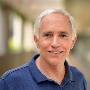
presented by Dr David Ress, Baylor College of Medicine where he is the Technical Director of the Center for Magnetic Resonance Imaging.
Abstract: The human brain exhibits a close relationship between neuronal electrical activity and blood flow. The phenomena, functional hyperemia, is the basis for functional magnetic resonance imaging (fMRI). In particular, a brief (~2 s) period of electrical activity evokes a stereotypical fMRI response that is often called the hemodynamic response function (HRF). Our laboratory has developed experimental methods, consisting of an audiovisual stimulus together with a speeded task, to evoke reliable HRFs across the majority of cerebral cortex in a single, hour-long fMRI scanning session. The resulting spatial pattern of response amplitudes is very similar across subjects. In healthy young subjects, the temporal dynamics of the HRFs vary only modestly across cortex. However, the dynamics show significant changes with aging possibly associated with cardiovascular changes. We have also developed a simple model for the HRF based on a linear network approximation to the vasculature, coupled with a 1.5D convection-diffusion treatment of oxygen transport. The model provides a quantitative interpretation of the HRF in terms of blood flow and cerebral oxygen metabolism. Our goal is to utilize these experimental and modeling methods as a means to diagnose various forms of brain pathology.
Date and Time
Location
Hosts
Registration
-
 Add Event to Calendar
Loading virtual attendance info...
Speakers
 David Ress of Technical Director of their Center for Magnetic Resonance Imaging
Measurements and Modeling of the Hemodynamic Response in the Human Brain
The human brain exhibits a close relationship between neuronal electrical activity and blood flow. The phenomena, functional hyperemia, is the basis for functional magnetic resonance imaging (fMRI). In particular, a brief (~2 s) period of electrical activity evokes a stereotypical fMRI response that is often called the hemodynamic response function (HRF). Our laboratory has developed experimental methods, consisting of an audiovisual stimulus together with a speeded task, to evoke reliable HRFs across the majority of cerebral cortex in a single, hour-long fMRI scanning session. The resulting spatial pattern of response amplitudes is very similar across subjects. In healthy young subjects, the temporal dynamics of the HRFs vary only modestly across cortex. However, the dynamics show significant changes with aging possibly associated with cardiovascular changes. We have also developed a simple model for the HRF based on a linear network approximation to the vasculature, coupled with a 1.5D convection-diffusion treatment of oxygen transport. The model provides a quantitative interpretation of the HRF in terms of blood flow and cerebral oxygen metabolism. Our goal is to utilize these experimental and modeling methods as a means to diagnose various forms of brain pathology.
Biography:
David Ress was initially trained as an Electrical Engineer at the University of California at Davis (BSEE, 1980). He then made a transition to studies of plasma physics during graduate work at Stanford University that focused on magnetic fusion experiments at Lawrence Livermore National Laboratory (LLNL); this work led to a Ph.D. in EE in 1988. He continued his work in fusion physics in the laser-fusion program at LLNL. Dissatisfied by the increasing emphasis on weapons physics in the laser program, he returned to Stanford in 1997 to study neuroscience using the methods of electron tomography and functional magnetic resonance imaging (fMRI), with the latter eventually becoming the main focus of his work. During this time his research focused on the use of high-resolution and -sensitivity methods to investigate neuroscience issues in human vision. He became an associate professor at Brown University in 2005, where he played a key role in developing an MRI facility for their neuroscience program. He then moved to the University of Texas in 2007, where he assisted in the development of their new Imaging Research Center, as well as performing research in high-resolution fMRI and the modeling of cerebrovascular physiology and physics. He moved to Baylor College of Medicine in 2013, with continued work in similar topics, as well as serving as Technical Director of their Center for Magnetic Resonance Imaging.
Email:
Address:Human Neuroimaging Laboratory, Baylor College of Medicine, Houston, Texas, United States, 77030
Agenda
presentation: Measurements and Modeling of the Hemodynamic Response in the Human Brain
speaker:
Dr David Ress, Baylor College of Medicine where he is the Technical Director of the Center for Magnetic Resonance Imaging

