Medical ultrasound imaging: quantification and visualization of diseases
Medical ultrasound imaging is widely used for the diagnosis of various diseases because of its noninvasiveness and real-time performance. However, due to the ultrasound-specific phenomena, it is difficult to understand what is visualized on ultrasound images without the knowledge and experience of the examiner. To overcome this difficulty, many quantification techniques have been developed. This presentation will outline the factors that make ultrasound images difficult to understand. I will then describe our recent work to quantify and visualize liver diseases based on the statistics-based analysis of ultrasound echo signals.
Date and Time
Location
Hosts
Registration
-
 Add Event to Calendar
- Carleton University
- 11 Colonel By Drive
- Ottawa, Ontario
- Canada K1S5B6
- Building: Canal Building
- Room Number: 3101
- Contact Event Host
- Co-sponsored by CU@EMBS
Speakers
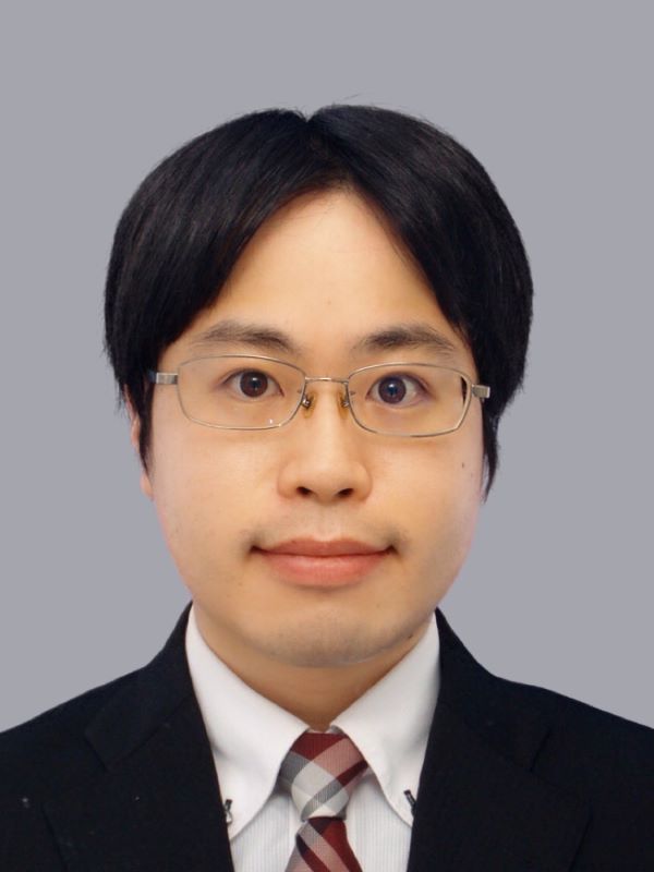
 Dr. Shohei Mori
Biography:
Shohei Mori is an Assistant Professor at Graduate School of Engineering, Tohoku University in Japan. He received the B.S., M.S., and Ph.D. degrees in engineering from the Tokyo Institute of Technology in Japan, in 2013, 2015, and 2017, respectively. His research interests include ultrasound tissue characterization based on the echo envelope statistics and ultrasonic measurements of organ dynamics for cardiovascular diseases.

